How are Microscopes Used in IVF, ICSI and IMSI?
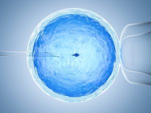 Assisted reproductive technologies (ART) refer to a variety of medical procedures and technologies used to help individuals or couples become pregnant. ART involves the manipulation of human eggs, sperm, or embryos in a laboratory setting to help overcome infertility or genetic problems. Some common types of ART include in vitro fertilization (IVF), intracytoplasmic sperm injection (ICSI) and intracytoplasmic morphologically selected Sperm Injection (IMSI). These technologies offer the possibility for individuals or couples who have difficulty conceiving naturally.
What are IVF, ICSI and IMSI?
IVF is the process of fertilization occurs outside the woman’s body in a laboratory setting. It involves stimulating the ovaries to produce multiple eggs, retrieving the eggs, fertilizing them with sperm in a laboratory dish, and then transferring the resulting embryos back into the woman’s uterus.
ICSI is a specialized form of IVF where a single sperm is directly injected into an egg to facilitate fertilization. It helps overcome fertility problems such as low sperm count, poor motility or abnormal morphology.
IMSI is an advanced version of ICSI where sperm are selected based on their morphology using high magnification microscopy. By using this advanced imaging technology, the most structurally sound sperm can be identified and selected to inject into the egg.
How are Microscopes Used in IVF, ICSI and IMSI?

Ovarian Stimulation Monitoring: Microscopes are used to monitor the growth and development of ovarian follicles during ovarian stimulation.
Egg Retrieval: When eggs are collected from a woman during IVF, microscopes are used to view the ovaries and guide the retrieval needle to safely and efficiently collect the eggs.
Sperm Selection: Microscopes can assess the morphology (shape), quantity, and motility of sperm. It is helpful in the selection of the most viable sperm for fertilization. Microscopes with high magnification are utilized to choose a single sperm for injection into the egg. In the case of IMSI, high magnification microscopes with up to 6000x magnification are employed for highly detailed sperm selection.
Embryo Assessment: After fertilization, microscopes help in assessing the quality of embryos based on cell division and morphology.
Embryo Selection: The best quality embryos are selected for transfer using microscopic evaluation.
Embryo Transfer: ln some cases, microscopes assist in the transfer of embryos into the uterus.
What Type of Microscopes are Used for IVF, ICSI and IMSI?
Assisted reproductive technologies (ART) laboratories require different microscopes to complete a variety of gamete (egg and sperm) selection and clinical procedures. Processes such as oocyte collection, embryo handling, and micromanipulation require various types of microscopes. The most common are high end microscopes with phase contrast attachments, inverted microscopes and stereo microscopes.
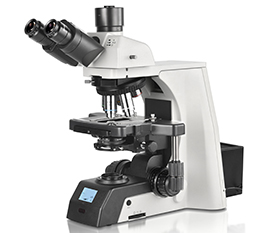
- High-End Upright Biological Microscope with Phase Contrast Attachment:
These microscopes are widely used to assess the morphology and structure of eggs. With the phase contrast attachment, the imaging contrast of the transparent specimen can be enhanced, enabling a clearer view of the details of the egg. This is essential for selecting healthy eggs for subsequent fertilization and culture. They can also accurately assess the morphology and motility of sperm and select the sperm with the most fertility potential.
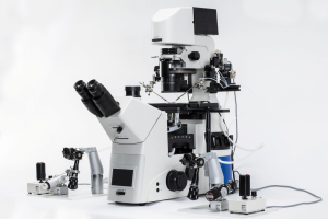
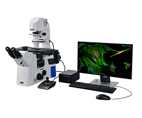
- Inverted Biological Microscope with Micromanipulators:
Inverted microscope is an important tool for microinjection. For the microinjection process, a single sperm is injected into the egg, prompting fertilization to occur. The design of the inverted microscope allows the sample to be viewed from the bottom, providing good field of view and control for precise injections. The inverted microscope required has to be equipped with the following features: a heated stage with 37°C, precise optics, micromanipulators, and injectors. Micromanipulators control the injection needle through tiny movements in microinjection technology to ensure the accuracy and precision.
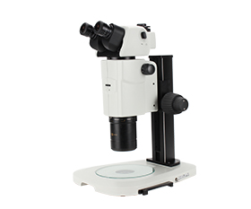
- High-End Stereo Microscope:
Stereo microscope provides a 3-dimensional or stereoscopic image, making it easier to observe the complete field of view. They are utilized for assessing the visual characteristics of ova, including size, shape, and surface details. During the pre-processing of egg in the IVF procedure, stereo microscopes aid in the precise removal of surrounding cellular tissues. In embryo assessment, stereo microscopes provide imaging that assists in the selection of embryos with optimal potential for successful implantation.
IVF, ICSI and IMSI Solutions:
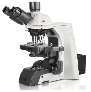
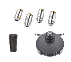 BS-2081 Research Biological Microscope
BS-2095 Inverted Biological Microscope with Micromanipulator
BS-3080 Parallel Light Zoom Stereo Microscope
The resources are collected and organized on the Internet, and are only used for learning and communication. If there is any infringement, please contact us to delete.


