


Scanpro2-6 Digital Pathology Section Scanner
Introduction
The Scanpro2-6 Digital Pathology Section Scanner consists of an imaging system, a micro-optical system, a control system, a scanning camera, a precision scanning platform and application software.
Details
Overview
Packaging & Delivery
Packaging Details:Strong Carton with Polyfoam Protection
Port:Beijing
Lead Time:Within 2-4 weeks after receiving payment
Introduction
The Scanpro2-6 Digital Pathology Section Scanner consists of an imaging system, a micro-optical system, a control system, a scanning camera, a precision scanning platform and application software.
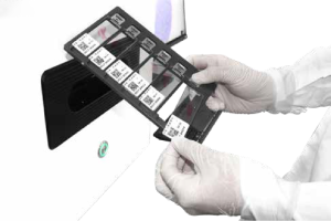
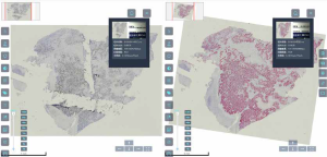
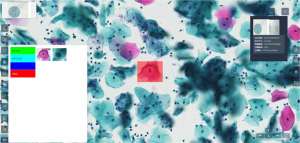
The users only need to complete the loading of slices, and the system can automatically complete slice loading, label recognition, area recognition of pathological slices, autofocus and automatic scanning. Finally, the pathological slices are presented in the form of digital slices on the computer display. Users can preview the digital slices globally, scale the key area, annotate and measure. The digital slices can be saved or transmitted separately for teaching or remote diagnosis.
Feature
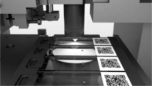
1.High success rate of scanning
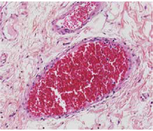
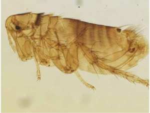
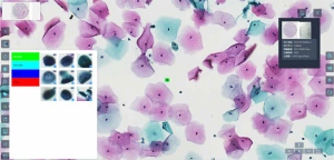
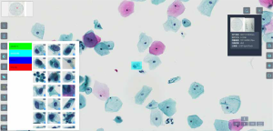
The Scanpro2 series pathological section scanner has excellent adaptability and can accurately scan all types of pathological sections. The Scanpro2 series can provide high-quality scanning results, whether it is lighter colored sections, uniquely shaped isolated pathological sections, TMA tissue chips, puncture tissue sections, or 2-inch/4-inch large tissue sections.

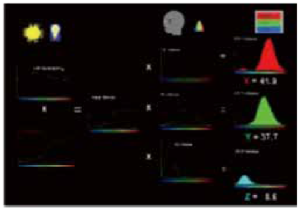
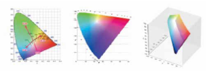
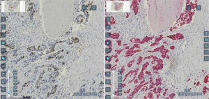
2.High fidelity color restoration technology
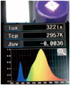
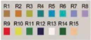
High fidelity color restoration technology enhances image brightness and color authenticity through advanced image processing. Combined with a prism spectrophotometer, ensure accurate reproduction of color when pathologists observe slices.
3.High speed scanning
The pathological section scanner is equipped with advanced high-speed scanning technology, which can efficiently scan 15mm*15mm sections in just 20 seconds, greatly improving the speed and accuracy of pathological diagnosis.
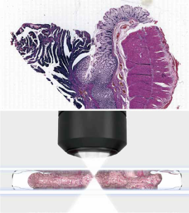
4.Depth of field fusion technology
By using high-precision multi-layer scanning technology, the projection image stack can be processed through specific fusion rules. This process involves maximizing the extraction of clear focal information from the projected images at each position, and ultimately merging this information into a comprehensive and clear image.
5.No stuck, no broke
By using high sensitivity reflective sensors, magnetic grating positioning and a dedicated glass slide sample loading mechanism, the problems of stuck, falling and broke of the slides have been effectively solved. In addition, it also utilizes precise linear platforms and magnetic levitation technology to ensure the safety of samples during the scanning process.

6.Microscope simulation film reading function
Supports simulating the viewing field under the microscope eyepiece, allowing for free switching of different objective magnification and rapid movement of the platform in the X/Y direction.
7.Energy conservation
Automatic shutdown: Once the scanning task is recognized as completed, the device will automatically enter the shutdown process without the need for manual operation, saving time and reducing operational steps.
Energy saving mode: In automatic shutdown mode, the device will shut down all non-essential energy consuming modules, such as light sources, sensors, etc., to maximize energy savings.
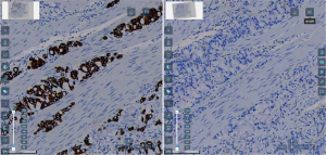
8.Multimodal comparison
9.Auxiliary analysis
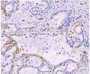
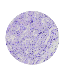
Supporting TCT and IHC assisted quantitative analysis (PD-L1, Ki67, etc.) for scientific research.
Specification
|
Item |
Specification |
Scanpro2-6 |
Scanpro2-50 |
Scanpro2-200 |
|
Maximum Loading Capacity |
6 slices |
● |
|
|
|
50 slices |
|
● |
|
|
|
200 slices |
|
|
● |
|
|
Supported Slides Specification |
1 inch, 2 inches, 4 inches |
● |
|
|
|
1 inch |
|
● |
● |
|
|
Objective |
Infinite Plan Super Apochromatic Objective 20X/ NA 0.8 |
● |
● |
● |
|
Scanning Magnification |
20X/40X |
● |
● |
● |
|
Focusing |
Autofocus. It can also be manually set |
● |
● |
● |
|
Sample Type |
Bright field slides such as HE stained slider, immunohistochemistry stained slides, frozen section stained slides, special stained slides, immunocytochemistry stained images, etc. |
● |
● |
● |
|
Slide Loading Type |
Manual slide loading |
● |
|
|
|
Automatic slide loading |
|
● |
● |
|
|
Scanning Method |
Line scanning |
● |
● |
● |
|
Scanning Camera |
4K line scanning camera, up to 66kHz line frequency |
● |
● |
● |
|
Scanning Area |
Automatic recognition & Manual setting, can be set according to different user needs |
● |
● |
● |
|
Scanning Method |
Bright field scanning: fully automatic scanning / manual custom area scanning |
● |
● |
● |
|
Scanning Resolution |
0.38u m/pixel (20x), ≤ 0.19 m/pixel (40x), collecting high-precision and high-quality images at the research level, achieving full information scanning of slices |
● |
● |
● |
|
Barcode Recognition |
Automatically scan the barcode for the slides identification and manage the scanned slides |
● |
● |
● |
|
Z-Stack Flight Focus Function |
Flight focus range 0-1.6mm |
● |
● |
● |
|
Scanning Speed |
20X scanning speed ≤25s (15*15mm) |
● |
● |
● |
|
Workstation |
Processor: Intel i7 and above |
● |
● |
● |
|
Memory: 32GB and above |
● |
● |
● |
|
|
Hard disk: 3TB |
● |
● |
● |
|
|
Display: 23.8 inches, 2k resolution, 1 unit |
● |
● |
● |
|
|
System software: Windows 10 |
● |
● |
● |
Note: ● Standard Outfit, ○ Optional


