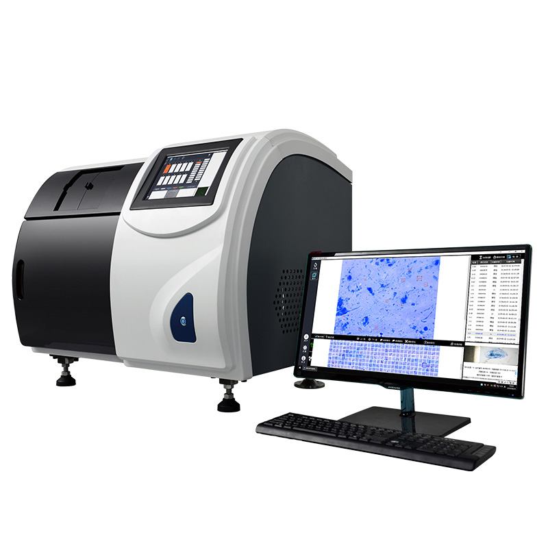
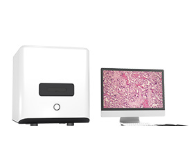
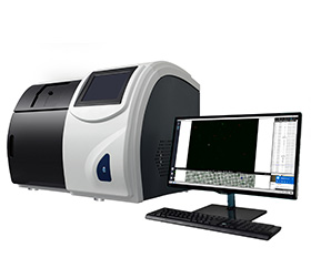
Scanpro 3-200FL Fluorescence Mycobacterium Tuberculosis Microscopy Scanning System
Introduction
The Scanpro3-200FL Fluorescence Mycobacterium Tuberculosis Microscopy Scanning System uses high-precision digital microscopy system to automatically acquire and preserve sputum smear image acquisition, intelligent recognition analysis and processing the image, combined with advanced artificial intelligence algorithm, the equipment give negative and positive grade to determine the sputum smear.
Details
Overview
Packaging & Delivery
Packaging Details:Strong Carton with Polyfoam Protection
Port:Beijing
Lead Time:Within 2-4 weeks after receiving payment
Introduction
Mycobacterium tuberculosis (M. tuberculosis), commonly known as tuberculosis, is the pathogen causing tuberculosis. Can invade all organs of the body, but the tuberculosis is the most common. Tuberculosis is still an important infectious disease. According to WHO statistics, about one out of every three people in the world is infected with Mycobacterium tuberculosis.
At present, the checkup of tuberculosis by medical institutions mainly depends on the traditional microscope, and using manual sputum smear. The traditional microscope is still facing following issues: Low efficiency, high undetected rate, big difference in diagnosis, difficult referral, sharing data cannot be saved, high work intensity and many other defects. Manual sputum smear microscopy inspection mode needs to be gradually replaced by automated equipment.
Scanpro3-200FL Fluorescence Mycobacterium Tuberculosis Microscopy Scanning System uses high-precision digital microscopy system to automatically acquire and preserve sputum smear image acquisition, intelligent recognition analysis and processing the image, combined with advanced artificial intelligence algorithm, the equipment give negative and positive grade to determine the sputum smear. Microscopy for Mycobacterium tuberculosis can be used for Ziehl-Neelsen acid-fast (antacid), auramine O staining (fluorescence).
Feature
1.Newly designed optical component system
A newly designed optical component system for automatic scanning of Mycobacterium tuberculosis, not a microscope modified system. Using custom optics scanning, the equivalent of 100 times the lens of the manual microscopy to 1600 standard field of view, the observation area covers the standard sputum area of 32%. The lens does not require refueling, do not need daily cleaning Maintenance, long lens life.
2.Smart scanning
Fully intelligent continuous scanning, one-click operation, 24 hours unattended, greatly improving work efficiency, and automatically cutting off power after scanning.
3.High-flux scanning
Supporting up to 200/50 sheets of smear at once, each fine scanning takes only about 3 minutes, and digital images can be permanently saved. During normal operation, the system automatically completes the loading and retrieval of smears. Supporting the urgent scanning of temporary smears. Supporting simultaneous scanning and reading of smear. Scan results can be customized for retrieval, statistics, etc.
4.Simple operation
Just put the processed smear into the system slide box at once, click “Start” in the computer. The Process can be stopped at any time with one key and emergency smears can be ready for insertion.
5.Intelligent identification
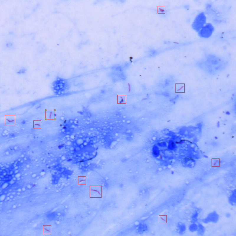
Powerful intelligent identification and analysis software, automatically identifies Mycobacterium tuberculosis, forms auxiliary diagnostic recommendations, and can customize bacterial model libraries according to different hospitals.
6.Information sharing
It can achieve information sharing with hospital information systems, as well as between doctors and patients. The smears can be viewed and discussed remotely via the Internet or local LAN, so that cross hospital diagnostic communication is no longer a problem.
7.System work flow
Process: Automatically scan images and digitally save. Image intelligent set up and analyze. Sputum smear negative / positive grade recommendations are automatically given. Auxiliary diagnosis.
Functions: Scan results pre-screening function. Urgent slice scan check function. Scan results custom statistics function. Binocular microscope inspection function. Target positioning / visual auto function.
Ziehl-Neelsen Acid-fast Staining
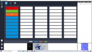
Scanning interface
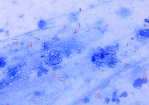
Intelligent identification
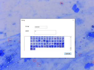
Diagnostic report
Auramine O staining
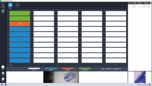
Scanning interface

Intelligent identification
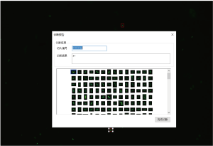
Diagnostic report
Specification
|
Item |
Specification |
Scanpro 3-50 |
Scanpro 3-200 |
Scanpro 3-50FL |
Scanpro 3-200FL |
|
Scanning Optical System |
A newly designed optical component system specifically designed for automatic scanning of Mycobacterium tuberculosis, without the need for refueling during scanning |
● |
● |
|
|
|
A newly designed professional fluorescence optical component system specifically designed for automatic scanning of Mycobacterium tuberculosis |
|
|
● |
● |
|
|
Manual Review Device |
Built-in integrated manual assisted lens inspection and review device, equipped with high eyepoint wide field eyepieces |
● |
● |
● |
● |
|
Illumination System |
Professional LED illumination system for microscopic observation, long lifespan, no flicker, energy-saving and environmental-friendly |
● |
● |
● |
● |
|
Maximum Loading Capacity |
50 slices |
● |
|
● |
|
|
200 slices |
|
● |
|
● |
|
|
Integrated slide box, supporting single slide urgent scanning, supporting scanning, loading, and reading at the same time |
● |
● |
● |
● |
|
|
Scanning motion mechanism |
The fast focusing mechanism with linear motor and magnetic levitation has good stability, low mechanical wear and long life |
● |
● |
● |
● |
|
Objective |
Infinity Plan Apochromatic Objective 20X, NA 0.5, WD=2.0mm |
|
|
● |
● |
|
Infinity Plan Apochromatic Objective 40X, NA 0.75, WD=0.74mm |
|
|
● |
● |
|
|
Infinity Plan Apochromatic Objective 60X, NA 0.9, WD=0.26mm |
● |
● |
|
|
|
|
Camera |
Professional camera with ultra-high resolution, 7 mega-pixels, 3200*2200, target size: 1.1” |
● |
|
|
|
|
Professional camera with ultra-high resolution, 4 mega-pixels, 2048*2048, target size: 1” |
|
● |
|
|
|
|
Professional fluorescent camera with high brightness and ultra-high resolution, 4 mega-pixels, 2048*2048, target size: 2/3” |
|
|
● |
● |
|
|
Label Recognition |
Panoramic camera, supporting automatic recognition of the entire slides and its label |
● |
● |
● |
● |
|
Scanning Time |
1-4 minutes for each slice, depending on the scanning area and smear quality |
● |
● |
● |
● |
|
Electrical Equipment Requirements |
Meeting medical device requirements |
● |
● |
● |
● |
|
Others |
Supporting depth of field extension, multi-layer scanning, coating area recognition, automatic/manual control of scanning area and scanning position, etc. |
● |
● |
● |
● |
|
Slide Loading Type |
66cm(L)*50cm(W)*67cm(H) |
● |
|
|
|
|
86.5cm(L)*67cm(W)*71.4cm(H) |
|
● |
|
|
|
|
66cm(L)*50cm(W)*67cm(H) |
|
|
● |
|
|
|
86.5cm(L)*67cm(W)*71.4cm(H) |
|
|
● |
● |
cNote: ● Standard Outfit, ○ Optional


