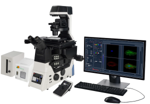
BCF296 Laser Scanning Confocal Microscope
Introduction
As life science research demands deeper and broader analysis of tissues, organs and model organisms, efficient imaging solutions have become pivotal for scientific breakthroughs. Capturing images of such large samples requires expanding imaging ranges and reducing acquisition times to detect more accurate intracellular reactions. Following the legacy of its commercially successful predecessors, the BCF295 confocal microscopes, we proudly announce the triumphant upgrade completion of our all-new generation confocal microscope, the BCF296, specifically designed to meet the stringent requirements of modern biological research for high-throughput and high-quality imaging.
Details
Overview
Packaging & Delivery
Packaging Details:Strong Carton with Polyfoam Protection
Port:Beijing
Lead Time:Within 2-4 Weeks after Receiving Payment
Introduction
Introduction
As life science research demands deeper and broader analysis of tissues, organs and model organisms, efficient imaging solutions have become pivotal for scientific breakthroughs. Capturing images of such large samples requires expanding imaging ranges and reducing acquisition times to detect more accurate intracellular reactions. Following the legacy of its commercially successful predecessors, the BCF295 confocal microscopes, we proudly announce the triumphant upgrade completion of our all-new generation confocal microscope, the BCF296, specifically designed to meet the stringent requirements of modern biological research for high-throughput and high-quality imaging.
This upgrade not only marks significant advancements in optical systems, resolution, and acquisition speeds, but also introduces profound optimizations in intelligent operation and motorized design, transforming intricate experimental procedures into effortless tasks, thereby fulfilling the stringent demands of cutting-edge research across disciplines.
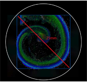
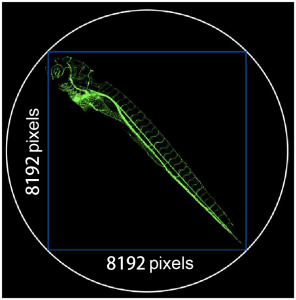
The BCF296 is equipped with an industry-leading 25mm field of view, effortlessly encompassing extensive sample areas for seamless large-scale imaging. With a scanning resolution of 8192*8192 pixels, it achieves vivid and precise imaging even at low magnifications. Its remarkable high-speed imaging capability of up to 60 fps (at 8*256 pixels) not only accommodates broad field observations but also delivers high-resolution imagery within a single frame, thereby gathering more data faster in each image.
Larger fields of view, enhanced resolution, and increased speed translate to superior image quality, clearer contrasts, and truer colors. This empowers you to delve deeper into the nanoscale universe within cells, interpreting cellular reaction processes more intuitively and accurately. It propels the boundaries of your scientific research forward, continually exploring uncharted territories in the realm of life sciences.
Feature
- To extract more data from the sample
The BCF296 confocal microscope boasts an industry-leading 25mm field of view, enabling it to capture images of large samples in a single scan and offering 1.5 times the data throughput compared to previous generations. Coupled with a scanning resolution up to 8192*8192 pixels, it is designed to meet the escalating demands in life science research for analyzing tissues, organs, and live model organisms, ensuring the acquisition of vital biological data with unprecedented detail and efficiency.
- Requires fewer total images for large and high-resolution image stitching
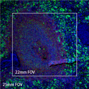
When paired with the BS-2096 inverted microscope, the BCF296 is capable of acquiring high-quality 25 mm field of view (FOV) confocal images. The large field of view reduces the number of images required for stitching large images and decreases image acquisition time, enabling uniform brightness, high efficiency, and high throughput imaging of large specimens.
22mm FOV large image stitching
25mm FOV large image stitching
- High-performance objectives for confocal imaging
The NIS series objectives boast high numerical apertures, long working distances, and exceptional chromatic aberration correction capabilities. Employing multi-layer coating technology, they deliver outstanding image quality and resolution. Not only do the NIS series objectives serve as an ideal complement to traditional optical microscopes, but they also play a pivotal role in confocal microscopy systems, enabling researchers to capture hitherto unseen fine structures and dynamic processes, thus facilitating in-depth exploration and visualization research of the microscopic world.
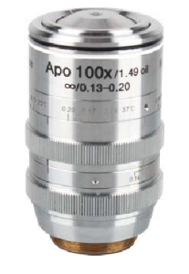
NIS Apochromat TIRF 100X Oil
Boasting unprecedented ultra-high resolution with a numerical aperture (NA) up to 1.49, this objective lens represents the pinnacle of the NIS series. It is meticulously designed to optimize spherical aberrations produced under varying imaging temperatures, namely 23 degrees Celsius and 37 degrees Celsius.
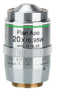
NIS Apochromat 20× C WI
The optimal choice for observing cultured cells and other specimens in culture medium, with a refractive index closely matching that of both the cells and the culture medium, thereby minimizing spherical aberration and light loss caused by refractive index differences to the greatest extent.
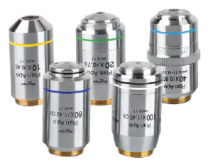
NIS Plan Apochromat series
Representing the apex of professional-grade objectives, with numerical apertures and working distances reaching new extremes, these lenses enable impeccable aberration correction across the entire field of view, offering imaging quality unparalleled by conventional objectives.
High-efficiency scanning heads and detectors
The BCF296 employs an internally integrated high-precision scanning galvanometer system within its scanning head, coupled with a continuously variable electric pinhole, enabling low noise, high contrast, and premium quality confocal imaging. With a scanning size of 8192*8192 pixels, it transcends the optical diffraction limit even when using low magnification objectives, achieving exceptional high-resolution sampling. This ensures the meticulous capture and reproduction of micro details.
- Faster scanning speed
Capable of high-speed imaging up to 60 frames per second (fps) at 8*256 pixels, reducing exposure time for samples under high light intensities and thereby significantly decreasing phototoxicity. This rapid imaging speed facilitates high-frequency data acquisition, enabling the capture of dynamic events and prolonged changes in samples, accurately and in real-time, fulfilling the complex and stringent imaging requirements of the life sciences domain.
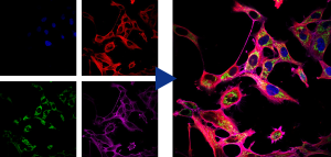
- Real-time four-channel synchronized imaging
The BCF296 confocal microscope incorporates cutting-edge four-channel fluorescence merging technology, empowering researchers to conduct real-time, synchronous, multi-channel precise fusion observations and captures. It allows for simultaneous detection and analysis of four distinct fluorescent labels within the same field of view, seamlessly switching and integrating multiple signals. Coupled with precise spectral unmixing imaging, this technology vividly and three-dimensionally reveals intricate, multi-layered information within samples, vastly enhancing experimental throughput and data accuracy.
- A powerful and integrated software platform for advanced analysis and visualization
The confocal microscope adaptation software achieves a high level of integration and control over the hardware devices of the confocal system and the core functions of the microscope. It seamlessly combines these controls with confocal imaging analysis, creating a high-performance, user-friendly, all-in-one experimental solution. Whether faced with complex application scenarios or specific research demands, the software ensures a seamless workflow experience for users through its outstanding integration and flexibility, freeing them from cumbersome microscope operations and allowing for a more focused approach on the essence of experiments and innovative exploration.
High-speed hardware control
It empowers users with unprecedented convenience in operation, effortlessly digitizing management and enabling precise control over multiple electric components within the microscope, such as objective lens switching, focusing, condenser lens changing, and fluorescence module transitions.
Objective magnification, light intensity and spectral control
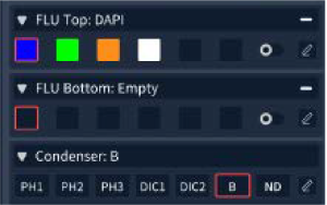
Fluorescence channel selection and observation mode control
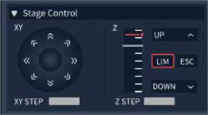
Stage control
Multi-dimensional imaging and image display
It is capable of memorizing custom observation modes and supports the combined use of X, Y, Z, λ, and T scanning functions. Equipped with a variety of flexible shooting modes, including multi-channel fluorescence imaging, time-lapse scanning, multi-position acquisition, Z-axis stacking, and panoramic stitching. These five modes can be freely combined according to the user’s actual needs, adapting to a wide range of complex and diverse experimental application scenarios.
Deconvolution
This feature allows for deblurring of two-dimensional images. It facilitates multiple iterations of deconvolution to eliminate the out-of-focus noise components, known as speckle noise, in confocal images. Additionally, 3D deconvolution is applicable to multidimensional images, further enhancing image clarity and resolution across multiple axes.
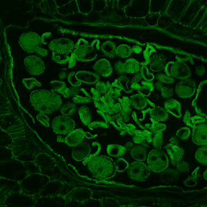
Before Deconvolution
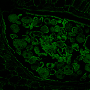
After Deconvolution
High-performance confocal microscopy platform
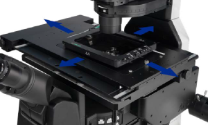
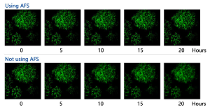
BS-2096 offers a powerful and flexible imaging solution, establishing a robust and highly expandable foundation for microscopic imaging within the BCF296 system. With its 25mm field of view design, it provides ideal observation conditions for large sample and high-throughput experimental research. Integrating various microscopical techniques such as brightfield, fluorescence, differential interference contrast, and phase contrast, users can freely opt for single-layer or dual-layer optical path configurations based on their specific experimental needs to achieve optimal imaging results. The Adaptive Focus Shift System (AFS) ensures precise focal plane positioning during continuous observations, thereby enabling stable, continuous, and clear recordings of cellular dynamic behaviors.
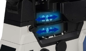
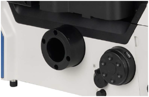
- High-speed electric motor control
The operation and switching speed of objectives, filter blocks, XY stage, and observation modules have been significantly enhanced, creating an effortless operating environment that enables researchers to focus on daily observations and image capture. A joystick for intuitive manipulation of the stage allows the microscope to become an extension of your eyes and hands, making it user-friendly and natural to operate.
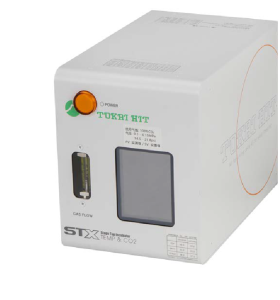
Living cell culture system
Specifically designed for precise live cell imaging, this system accurately regulates the microscope platform temperature, ensuring constant temperature, humidity, and CO2 concentration within the culture dish, thereby
providing an ideal cultivation environment for long term experiments.
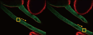
AFS ensures stable and reliable imaging performance
The BCF296 adopts an independent focusing design, minimizing the impact of other mechanical components on the Z-axis. It features a newly designed Adaptive Focus System (AFS), which intelligently eliminates focus drift. Whether paired with high-magnification objectives has large numerical apertures or utilized in conjunction with advanced imaging techniques such as super-resolution, confocal, or TIRF (Total Internal Reflection Fluorescence), the system consistently delivers crisp, sharp images. This design ensures the highest level of imaging stability and precision across a broad range of demanding applications in modern microscopy.
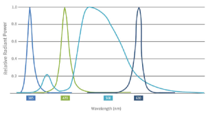
High specification LED fluorescent illumination system
The LED illuminator enables up to 4-channel LED illumination, offering high compatibility with commonly used fluorescent dyes in the market. It features concentrated excitation energy and high brightness, fulfilling the fluorescence imaging requirements of routine experiments. With instant-on capability, long service life, and no need for bulb replacement, it outperforms traditional mercury arc lamps in terms of reducing photobleaching and phototoxicity, making it particularly friendly to live cell samples. It is a sustainable, energy-efficient, and environmentally friendly microscope light source, ideal for low-carbon laboratory practices.
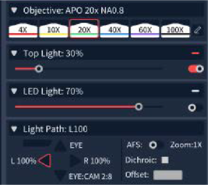
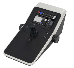
Interactive operation
BS-2096 innovatively incorporates a touch screen into its front panel, significantly enhancing user interface convenience and expandability of functions. It retains the traditional microscope knobs and buttons on both sides ensures intuitive control even in dark laboratory environments, allowing researchers to concentrate on the core of their experiments without being hindered by complicated operations. This design promotes an efficient and seamless microscopy observation experience.
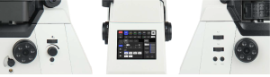
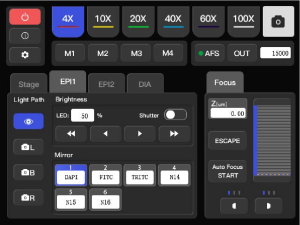
5.6inch touch screen
Control of components such as objectives, dual-layer/single layer fluorescence filter wheels, the condenser, light intensity, electric stage speed, electric Z-axis speed, host spectrometer ports, ESC exit, FN keys, and objective parfocality is achieved through the touch interface. It also provides real-time display of various statuses including objective magnification, transmitted illumination brightness, fluorescence wavelengths, output ports, XYZ positions, and movement speeds.
Large aperture observation optical system
Equipped with a large-aperture objective lens, it significantly increases light transmission, coupled with a spacious CMOS sensor, effortlessly enabling brightfield and fluorescence imaging across a vast field of view up to FOV25mm. This broader perspective captures more details, empowering you to comprehensively explore the microscopic world and have complete control over your scientific research endeavors.
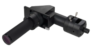
Large aperture reflective fluorescence illuminator
Specifically designed for a large field of view (FOV) of 25mm, this fluorescence imaging illumination apparatus features a high-power LED light box, delivering broadband, high-transmission illumination encompassing the ultraviolet spectrum. It is also compatible with large-aperture fluorescent filters, ensuring high signal-to-noise ratio fluorescent images for detailed and accurate observations.
Specification
|
Item |
Specification |
BCF296 |
|
|
Optical system |
Infinite optical system |
● |
|
|
Laser |
Laser 405 nm, 488 nm, 561 nm, 640 nm |
● |
|
|
Detector |
Standard detector, wavelength: 400-750nm, 4-channel PMT detector |
● |
|
|
Transmittance detector, single channel PMT detector |
○ |
||
|
Scanning head |
Maximum pixel size: 8192*8192 |
● |
|
|
Scan Mode |
Supports combined use of X, Y, Z, λ and T scanning functions |
● |
|
|
Pinhole |
Electric stepless adjustment |
● |
|
|
Confocal Field of View |
Square inscribed in a Φ25mm diameter circle |
● |
|
|
Eyepiece |
10X, adjustable diopter -5~+5 |
● |
|
|
Viewing Head |
Siedentopf binocular viewing head, 10-40° inclined, Interpupillary 47-78mm, eyepiece interface Φ30mm. |
● |
|
|
Objectives |
Apochromatic Plan Objectives |
4X/ NA=0.16, WD=17.0mm, cover slip=0.17mm |
○ |
|
10X/ NA=0.45, WD=4.0mm, cover slip=0.17mm |
● |
||
|
20X/ NA=0.75, WD=1.1mm, cover slip=0.17mm |
● |
||
|
(Wi) 20X/ NA=0.95, WD=1.1mm, cover slip=0.11-0.23mm |
○ |
||
|
40X/ NA=0.95, WD=0.21mm, cover slip=0.11-0.23mm |
● |
||
|
(Oil) 60X/ NA=1.42, WD=0.25mm, cover slip=0.17mm |
● |
||
|
(Oil) 100X/ NA=1.45, WD=0.13mm, cover slip=0.17 |
○ |
||
|
(Oil) 100X/ NA=1.49, WD=0.09-0.16mm, cover slip=0.13-0.19mm (23°C), 0.14-0.20mm (37°C) |
○ |
||
|
Nosepiece |
Motorized sextuple nosepiece with DIC expansion slot |
● |
|
|
|
Motorized sextuple nosepiece with AFS module |
○ |
|
|
Stage |
Motorized control (grating type): Stage size: 445mm*300mm, travel range: 130mm*100mm, maximum speed: 25 mm/s. Positioning Accuracy: 0.1 μm. Repeatability: 0.5 μm. Equipped with a universal stage plate, compatible with 35mm-65mm culture dishes and slides, optional plate holder adapter available. |
● |
|
|
Condenser |
7-hole turret condenser with electric control, NA0.55, WD26. 7 positions for phase contrast, Hoffmann phase contrast, DIC, ND filter. |
● |
|
|
Focusing System |
Electric Z-axis, travel distance 10mm, precision 0.02μm, repeatability 0.1μm. Three-speed focusing knob: 2μm/revolution, 40μm/revolution, 200μm/revolution. |
● |
|
|
Illumination System |
Transmitted Kohler illumination, 3W LED |
● |
|
|
Epi-Illumination: Widefield 4-band LED illumination. 6-Position electric fluorescence turntable (standard for B, G, U). Motorized Fluorescence Shutter. |
|||
|
Ports |
Beam splitter ratios: eyepiece 100% or left port 100% or right port 100% or eyepiece: right port=20:80. |
● |
|
|
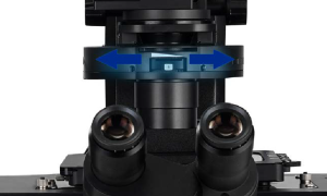
Intermediate Magnification |
Manual 1X, 1.5X switching |
● |
|
|
DIC Plate |
10X, 20X, 40X, 60X, 100X objective Inserts, compatible with the nosepiece turret |
○ |
|
|
Power control box |
X/Y/Z-axis electric control. Display objective magnification, fluorescence band and other states. Shortcut key settings. |
● |
|
|
Image Bit Depth |
16 bits |
● |
|
|
Camera |
Specialized camera |
● |
|
|
Software |
Software: Featuring confocal scanning capabilities, multidimensional imaging, and pre-set channel functions, this software supports both wide-field and confocal imaging modes. It offers comprehensive hardware control modules, built-in image filtering tools, and supports export to various image formats, providing users with advanced imaging processing and management options. |
● |
|
|
PC |
Computer workstation: a set of HP workstations or similar performance workstations. (1) HP Z840/CT Workstation/English OS Windows 7 64bit Professional Edition (2) CPU: Intel Xeon E5-26434C 3.30 10MB 1600 x 1 or similar performance (3) RAM: 32GB DDR -1600 ECC or similar performance (4) HDD: 1TB 7200 RPM SATA 1ST HDD or similar performance (5) 16X SuperMulti DVDRW SATA 1st ODD or similar performance (6) Display: 2PCS ≥20 inch LED backlit widescreen IPS LCD displays. |
● |
|
|
Expandable Functions |
Live Cell Culturing System Super-Resolution Structured Illumination Microscopy (SR-SIM) Module Dual-Layer Fluorescence Heightened Accessor |
○ |
|
Note: ● Standard Outfit, ○ Optional
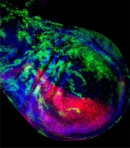
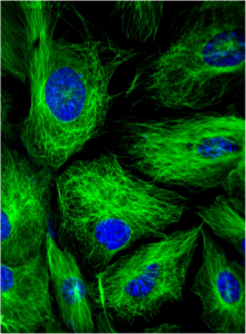
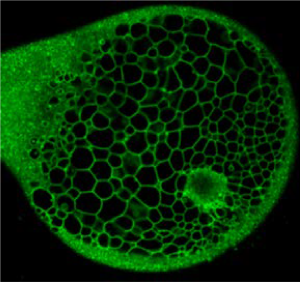
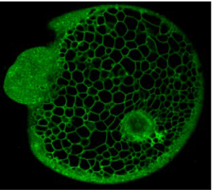
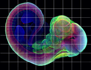
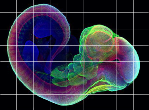
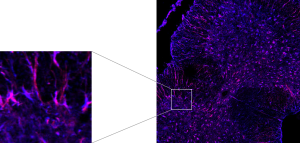
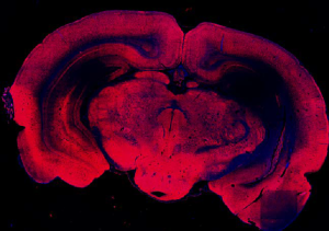
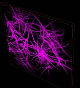
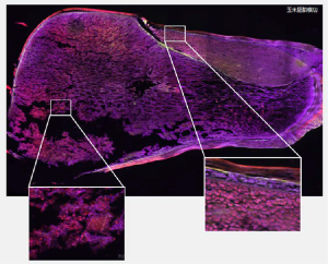
Sample Image