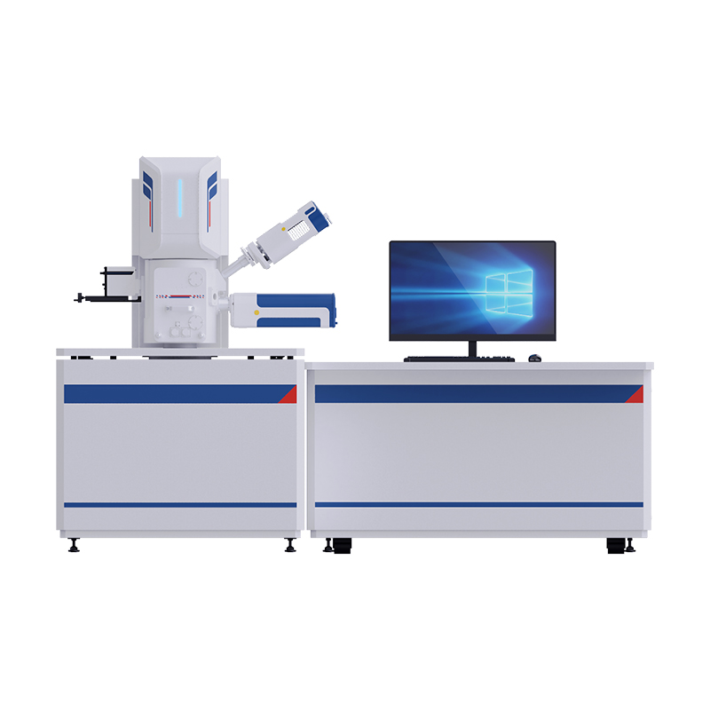
BSEM-801 Field Emission Gun Scanning Electron Microscope
Introduction
BSEM-801 is a Field Emission Gun Scanning Electron Microscope. It has excellent imaging quality capabilities. It has More than six preset extend interface for accessories such as WDS, EDS, EBSD, etc.
Details
Overview
Packaging & Delivery
Packaging Details:Strong Carton with Polyfoam Protection
Port:Beijing
Lead Time:Within 2-4 Weeks after Receiving Payment
Introduction
BSEM-801 is a Field Emission Gun Scanning Electron Microscope. It has excellent imaging quality capabilities. It has More than six preset extend interface for accessories such as WDS, EDS, EBSD, etc. It helps users to characterize specimens and explore the world of microscopic imaging and analysis.
Features
1.Specification
Resolution: 1.5nm@15KV (SE), 3nm@30KV (BSE)
Magnification: 8-800000X
Electron Gun: Schottky field emission gun
Accelerating Voltage: 0.2-30kV
Lens System: Multilevel high performance conical objective system
Objective Aperture: Three objective apertures adjustable outside the vacuum system, replacing without disassemble lens
Vacuum System: 1 set ion pump, 1 TMP, 1 mechanical pump. Specimen chamber vacuum better than 6.0E-4 Pa, electron gun vacuum better than 2E-7Pa, automatic vacuum control with vacuum interlock in order to avoid mis-operation
Detector: High vacuum secondary electron detector (SE), Integrated back scattering detector (BSE), IRCCD
More than six preset extend interface for accessories such as WDS, EDS, EBSD, etc.
2.5 axis motorized stage
Five-axis eucentric motorized stage: X=80mm, Y=50mm, Z=30mm, T=-5°~+70°, R=360°.
Max loadable sample: Ф175mm. Max height: 40mm.
Touching alarm and auto-stop, in case of mis-operation.
3.Image Processing
4096*3072 pixels display. Image Format: TIF, BMP, GIF, JPG, PNG. Automatic recording digital movie: (avi) function.
4.Work station
PC Workstation (Customized upon request). Image can be saved at hard or other storage medium, or printout.
Commercial workstation, Intel Xeon; 16G RAM+500G SSD+1TB graphics card, P1000 (independent graphics card -4G) +232 interface, 24” Monitor, wireless keyboard & mouse. Windows 10 operating system available in both Chinese and English language, images can be saved at external hard drive or print out.
5.Image Display
Frame averaging function, line average function, rich image hierarchy, rich image hierarchy, real-time display magnification, scale plate, acceleration voltage, grey curve.
6.Software Control
High voltage integrated commission, Automatic filament on/off, Potential shift regulation, Brightness adjustment, Electric to central adjustment, Automatic brightness, Contrast adjustment, Objectives adjustment, Auto focus, Magnification adjustment, Objective degaussing, Automatic astigmatism elimination, Selected area scanning mode, Electric rotation adjustment, Management of microscope parameters, Point scanning mode, Electron beam displacement adjustment, Real time display of scanning field size, Line scanning mode, Electron beam tilt adjustment, Gun lens adjustment, Surface scanning, Scanning speed adjustment, Multichannel input, High voltage power monitoring, Swing centering, Tape measure, etc.
Installation Requirements
1.Power supply requirements
Voltage: AC 220V ± 10%, 50Hz ± 1 Hz, standard sine wave.
It is not recommended to share the power supply line with the instrument for equipment with high power and large power consumption change.
Three power sockets needed for: Scanning electron microscope instrument body. Computer: AC 220V, 50Hz, 16A. Mechanical pump and air compressor: AC 220V, 50Hz, 16A.
Independent ground, make sure ground resistance less than 3Ω.
Power supply: AC 220V (+7%-10%), 50Hz (±1%). Air switch: 63A.
2.Environmental requirements for installation site
Temperature between 16-30°C is recommended. The relative humidity shall be less than 60%.
Recommend configuration: air conditioner, dehumidifier and other equipment that can ensure the temperature and humidity of the laboratory.
Noise: <68 DB.
The durability of the instrument operation: continuously working.
Vibration from any direction less than 3μm (P-P) at the instrument installed location.
Magnetic field from any direction less than 2×10⁻⁷T (2mG, P-P).
Instrument install location suggest first floor/ground floor stay away from railway, subway, main road as well as magnetic interference sources such as transformer equipment and high-frequency equipment.
Clean nitrogen (99.9%).
- To fulfill above environment condition is the key factor to reach the best performance of the instrument.
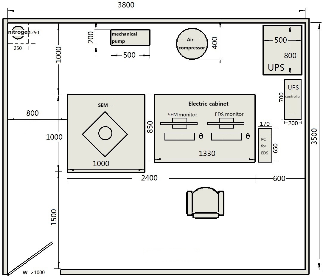
3.Instrument Dimension & Weight
Microscope body: 800mm*800mm*1480mm
Working table: 1340mm*850mm*740mm
Total weight: 450kg
Total power: 3kW
The floor bearing capacity should ≥ 500kg/m3, and it is recommended to place it on the first floor.
Specification
|
Item |
Specification |
BSEM-801 |
|
|
Electron Scanning Microscope |
BSEM-801 Field Emission Gun SEM |
● |
|
|
Five axis automatic specimen stage, X=80mm, Y=50mm, Z=30mm, T=-5°~+70°, R=360°. Max loadable sample: Ф175mm. Max height: 40mm. |
● |
||
|
Detectors |
SE detector |
● |
|
|
BSE detector |
● |
||
|
IRCCD |
● |
||
|
Control panel |
● |
||
|
PC |
● |
||
|
Air compressor |
● |
||
|
Consumable Items |
Field emission filament, installed in microscope |
● |
|
|
Sample cup, Φ13mm |
● |
||
|
Sample cup, Φ32mm |
● |
||
|
Carbon double-sided conductive tape, 6mm |
● |
||
|
Vacuum grease, not allowed through air transportation |
● |
||
|
Hairless cloth |
● |
||
|
Polishing paste, not allowed through air transportation |
● |
||
|
Sample box |
● |
||
|
Cotton swab, 100/bag |
● |
||
|
Oil mist filter |
● |
||
|
Tools |
Inner hexagon spanner, 1.5mm-10mm |
● |
|
|
Tweezers, length 100-120mm |
● |
||
|
Slotted screwdriver, 2*50mm, 2*125mm |
● |
||
|
Cross screwdriver, 2*125mm |
● |
||
|
Clean vent pipe, Φ10/6.5mm (out diameter/inner diameter) |
● |
||
|
Vent pressure reducing valve, output pressure -0.6*25 MPa |
● |
||
|
Internal baking power supply, 0-3A DC |
● |
||
Note: ● Standard Outfit, ○ Optional
Application
![]()
Silicon-based carbon material

Lithium iron phosphate
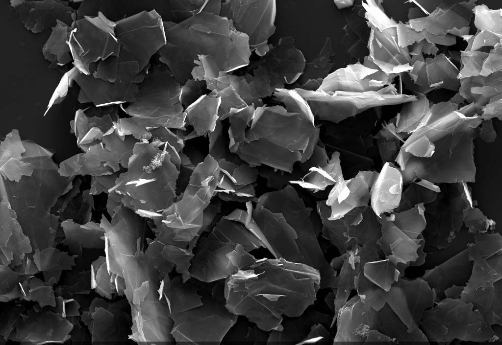
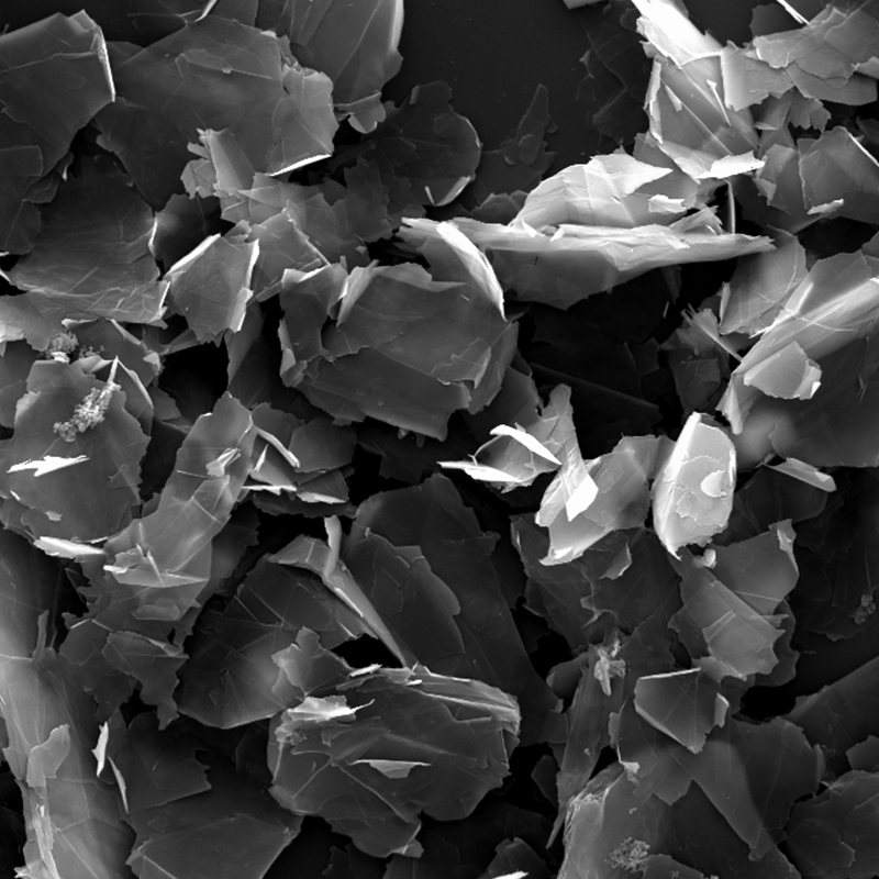
Graphene
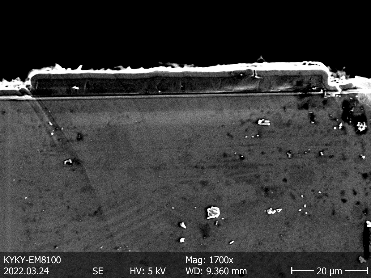
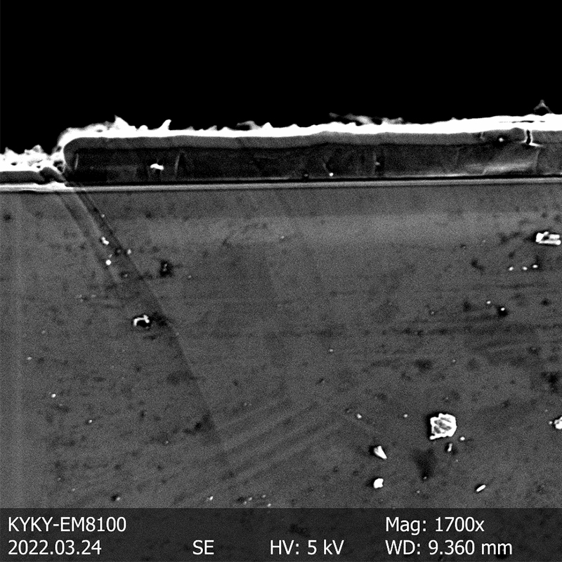
PN junction
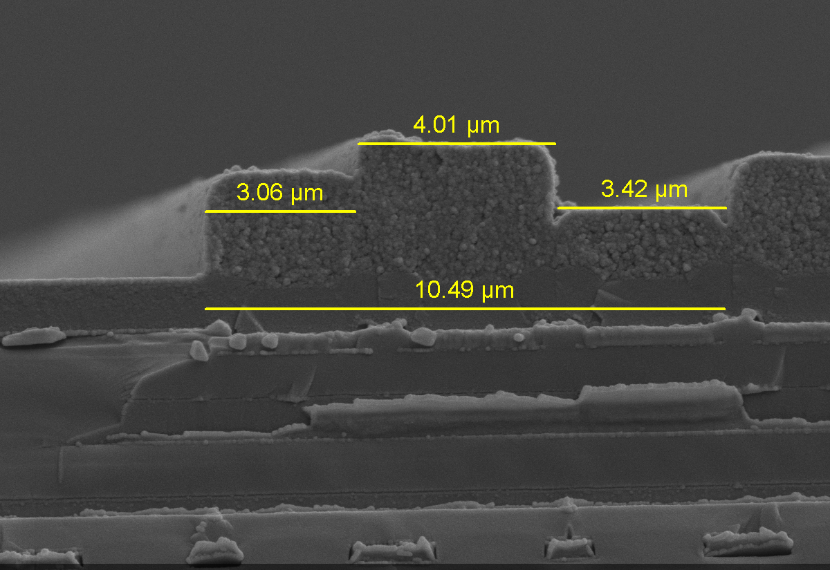
Etching layers for microchips
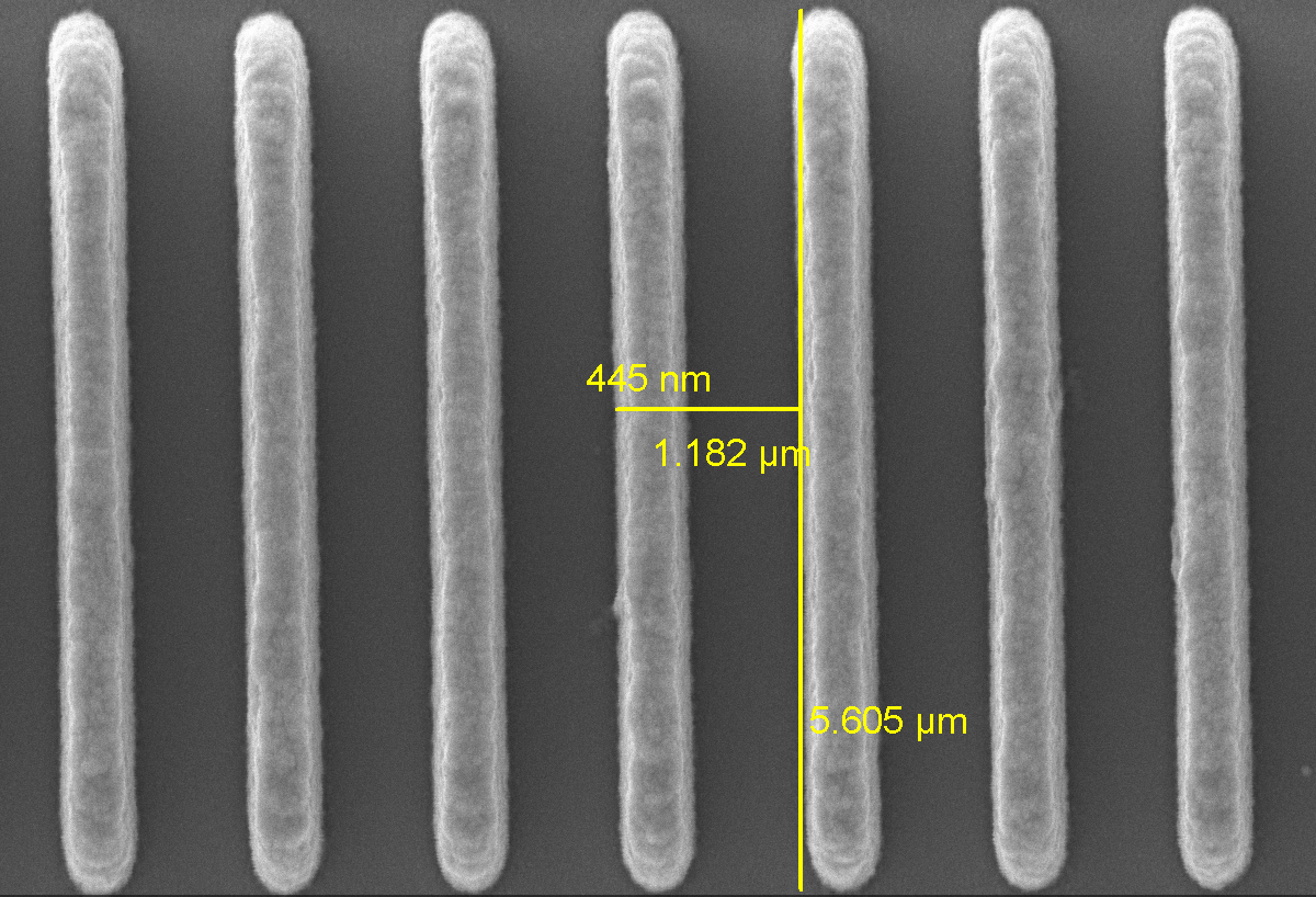
Line width measurement


